Some procedures have specific patient preparation requirements. This usually involves fasting for a set period. If there is specific preparation you need to follow, we will let you know when you make your appointment.
Please ensure you bring your referral, Medicare card and any previous films with you to your appointment.
When you arrive at our practice the reception team will register you for your procedure.
A friendly member of the GMI team will greet you, explain the procedure and gather specific clinical information, which will assist with your results, and enable us to perform the scan as accurately as possible.
On arrival you will be asked a series of in-depth safety questions to ensure it is safe for you to have this scan. It’s important that you answer these questions correctly.
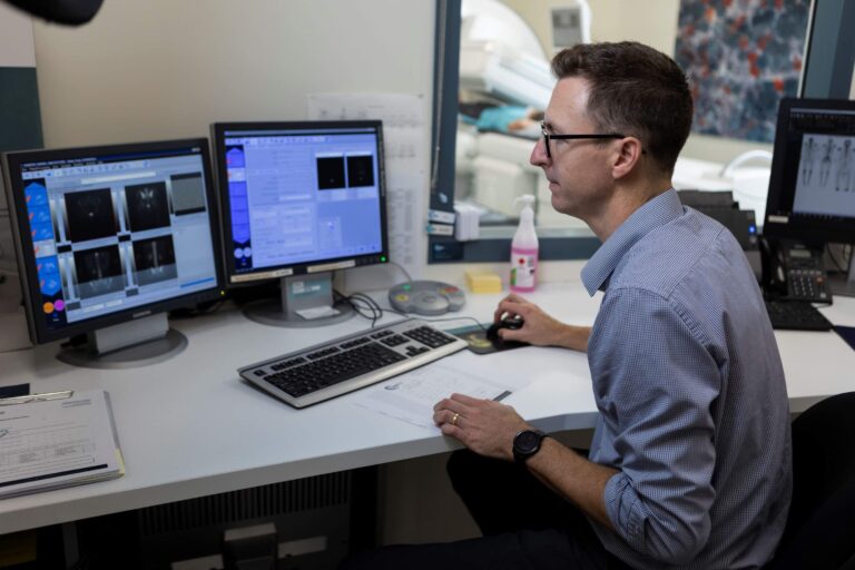
Once your images are complete, we process them using our computer. This allows us to provide clear images to assist in your diagnosis. Our doctor will complete the report and make the images available to your doctor.
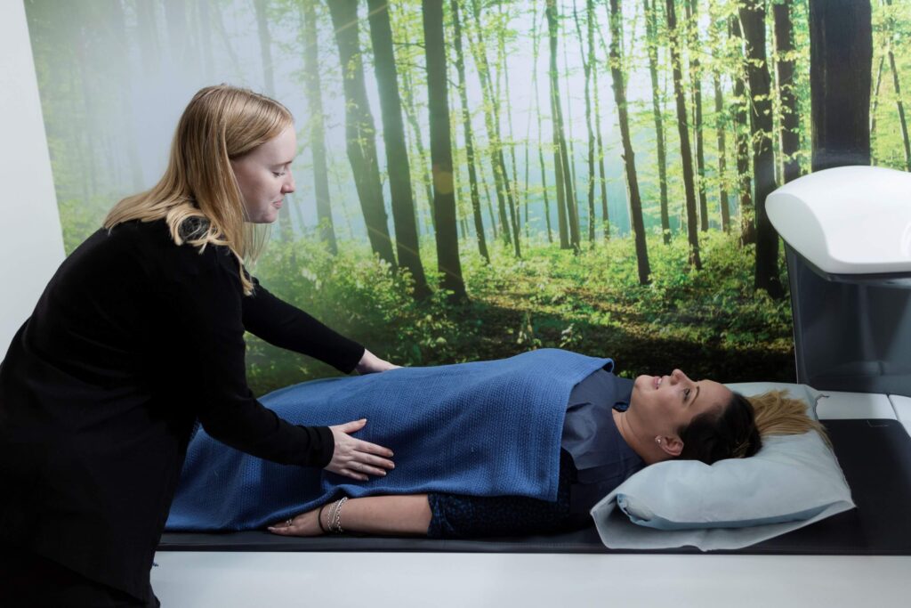
You will be asked to change into a gown and lie on the scanning table. Sometimes you may be required to sit beside the table as well for images of your extremities. The bone densitometry unit will then be moved into the correct position to take the image. The ‘arm’ of the machine will move along the body and take the measurement of your bone density.
The machine uses low energy, low dose x-rays that are emitted from the unit and pass through the body onto the detector. This detector converts the x-rays that interact with it into both a measurement and an image.
A typical bone density examination takes only 10 minutes to complete.
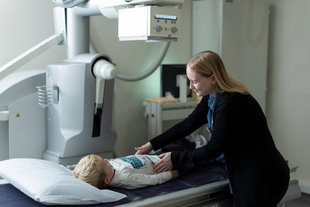
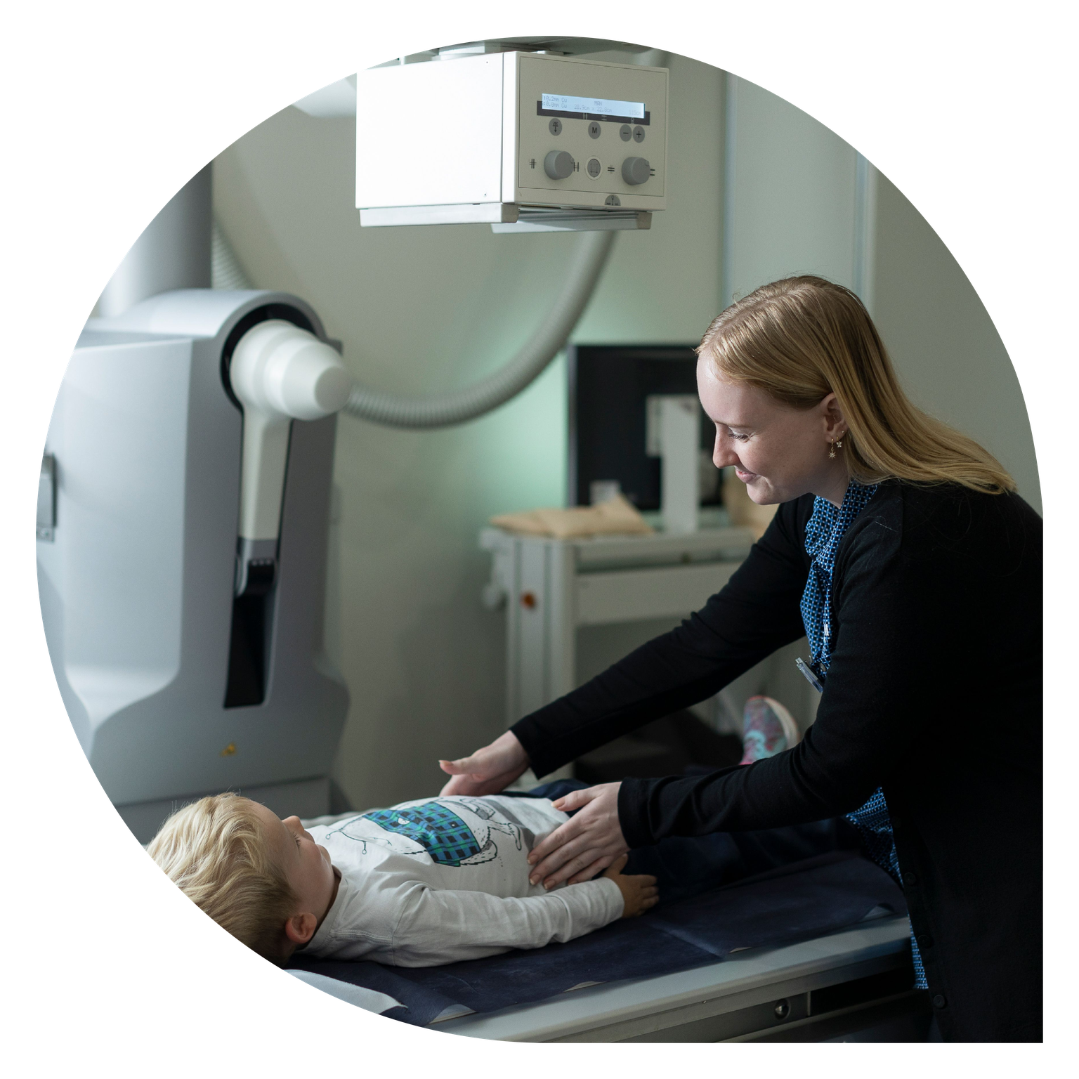
You will be asked to change into a gown and lie on the scanning table. The x-ray unit will then be moved into the correct position to take the image. Most screening or fluoroscopy studies require the administration of x-ray contrast (a liquid dye). This can be administered orally or injected into the area of concern. This contrast will highlight the area of interest and allow more accurate diagnosis.
The x-rays are emitted from the unit and pass through the body onto the detector. The detector converts the x-rays that interact with it into an image.
Screening examinations can vary greatly in time, typically less than 30 minutes, and sometimes just a few minutes. Our Siemens x-ray equipment is identical to the one shown here…
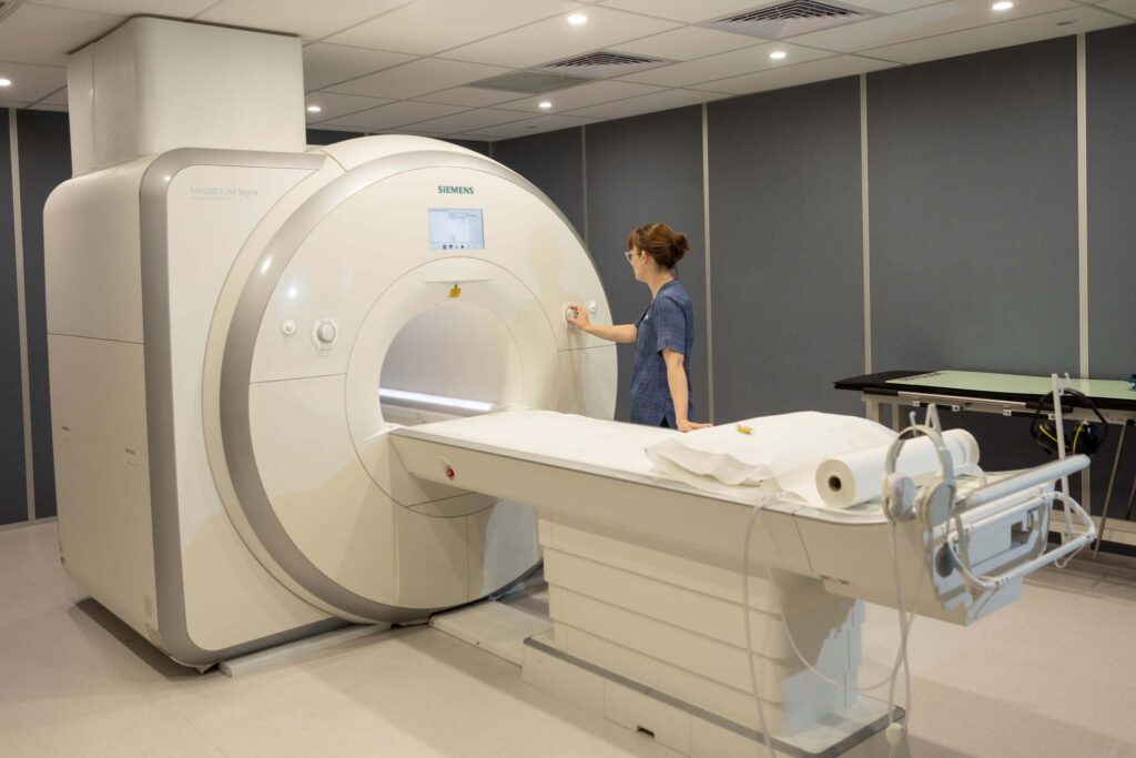
Before you enter the MRI room, you will asked to changed into a gown and remove all metallic objects.
You will be positioned on our MRI table (see background image) and some MRI coils will be placed around the part of your body to be imaged. The table will then slide into the MRI scanner and the test will begin.
The MRI can be noisy and you will be given headphones to help block out the noise. Our scanner has the ability to play music through the headphones, so you are welcome to bring some music with you to the MRI examination.
During the scan it is important to stay very still because MRI scans are very susceptible to motion.
Sometimes your scan may require an injection of gadolinium (a contrast dye). Gadolinium is very safe and should not affect you once you leave the MRI department.
You may feel a warm sensation during the scan, which is normal. If anything during your test concerns you please inform the radiographer immediately.
MRI scan times are variable, typically ranging from 20 minutes to one hour. Our staff will let you know how long it will take when you book your appointment.
Metal foreign bodies in the eyes can also prevent scanning. If you have ever had an injury to your eyes involving metal, please inform our staff. You may need to have an x-ray of your eyes to ensure no residual metal fragment is present.
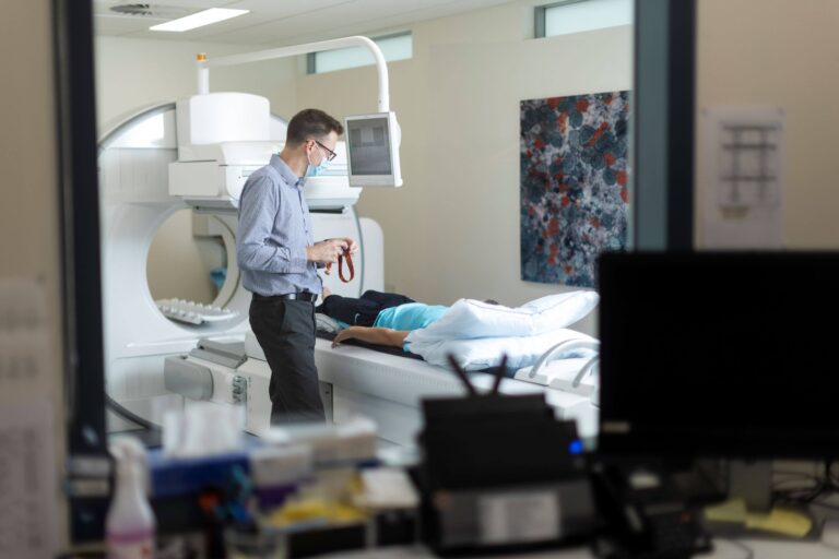
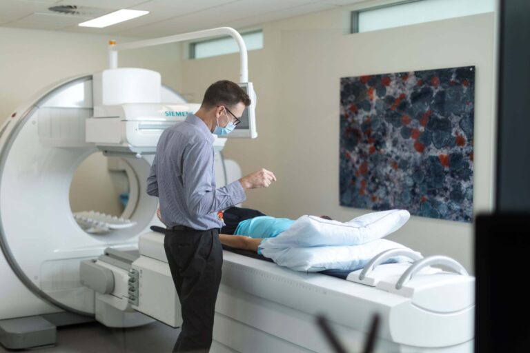
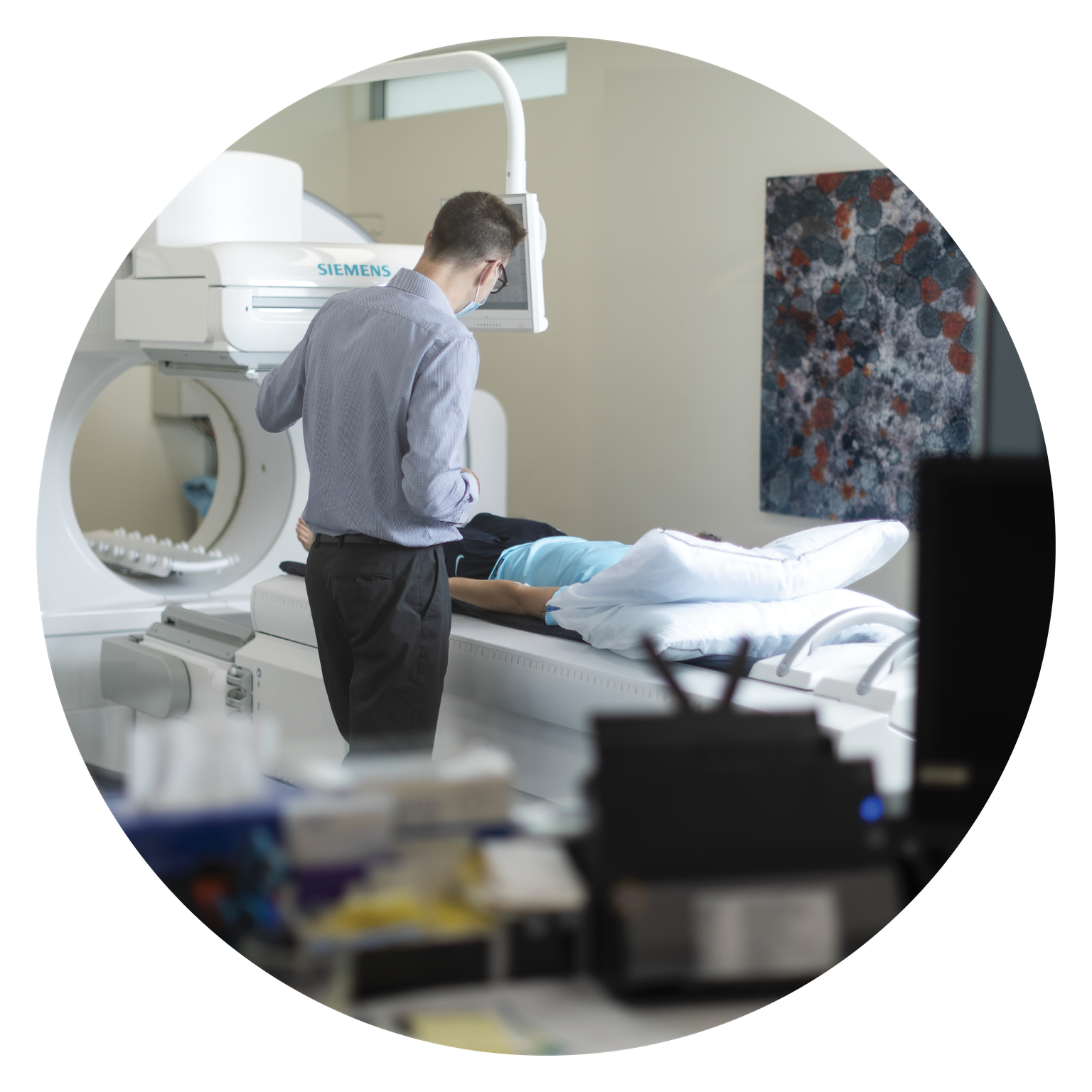
For nuclear medicine procedures a radiopharmaceutical is administered – this is usually given intravenously (into a vein in your arm), but for some scans it is administered orally (as a drink). This radiopharmaceutical follows specific functions within the body and allows us to take images of this functional information. We use a SPECT/CT Gamma Camera that utilizes the latest xSPECT technology.
Sometimes we need to acquire images when the radiopharmaceutical is administered and this can vary in duration. Other times we need you to return a few hours or days later to have the images taken. On some occasions we require both of the above. Any detailed information specific to your procedure or scan will be given to you before you arrive at our practice.
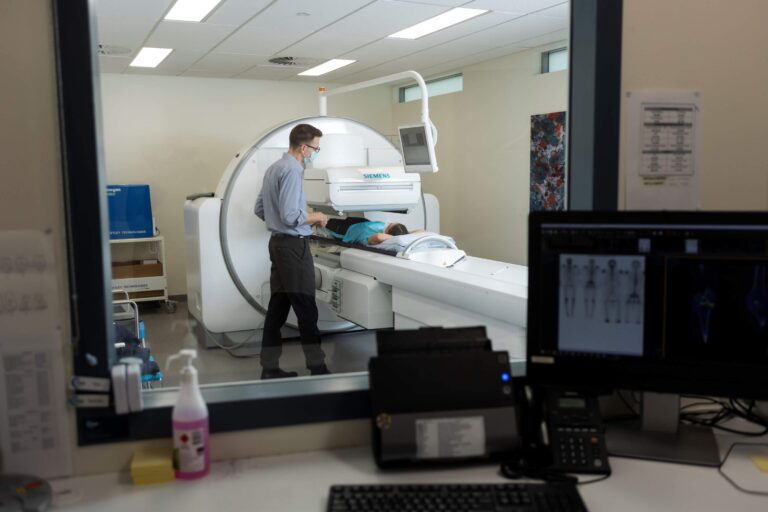
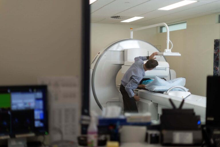
You will be asked to lie down on our scanning table as shown in the image. This will then move into the machine where the scan will occur. This process is painless and the machine is very quiet. Images can take from a few minutes to an hour depending on the type of scan.
On occasions the machine will rotate around you and acquire images in 3D – this is called SPECT. We may also have to take a low dose CT series that is reconstructed, fused with the 3D nuclear images and creates our SPECT/CT images.
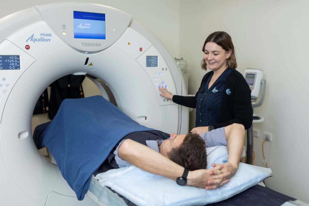
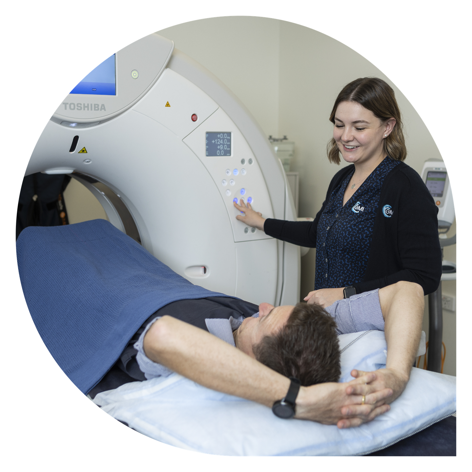
Some CT procedures require a liquid contrast (a type of dye). This can be administered orally – by drinking a fluid, or intravenously – via a cannula into a vein in your arm, or both. This contrast provides the scanner with much clearer visual images of certain structures within your body.
We use a 160 slice CT scanner which allows for fast scan times which lowers the patient dose of radiation.
You will be asked to lie on the scanning table that slides into the CT scanner, which looks like a large ring or donut. The x-rays are emitted from the ring and pass through the part of the body being scanned. The table moves in and out to allow the images to be taken. A computer detects the amount of radiation passing through the body and forms an image of the body part being scanned.
The scan usually takes no more than half an hour to perform. However, you may need to arrive in the x-ray department up to one hour before the examination if you have to drink contrast dye for the scan.
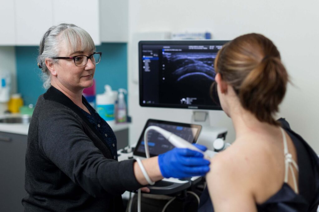
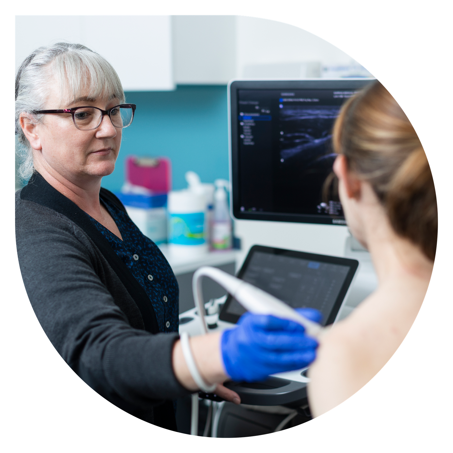
First you will change into a gown for the scan. You will then be asked to lie on the couch (bed) beside the ultrasound machine and expose the region to be examined. Your privacy will be respected throughout this process and the rest of your body will be covered during the procedure.
A clear gel is applied to the examination area– to allow the sound waves to be transmitted into the body. The ultrasound probe or transducer is placed onto the skin and moved across the surface to evaluate the structures below.
The scan is painless, although some pressure may be applied to improve the image. Please tell the sonographer if this becomes uncomfortable.
Examinations of the shoulder or other joints may require your cooperation in performing certain movements while scanning is in progress.
Doppler ultrasound uses special technology to detect blood flow in arteries or veins. During this process you will hear a ‘whooshing’ noise as signals from the blood flow are transformed into sound.
We may need to gently squeeze your calf when leg veins are examined.
Most ultrasound scans take 30 minutes to perform. However, some tests (especially doppler studies) may take up to two hours. Some other examinations or procedures may require more than one visit.