What is nuclear medicine?
Nuclear medicine encompasses the use of medical radioisotopes to take images of various parts or systems in the human body. It involves injecting tiny amounts of “radiotracers” into the body to assess the functions of various organs or body systems. It is most commonly used to assess the skeleton, heart, thyroid and kidneys. Unlike x-rays or CT scans (which send x-rays through your body) nuclear medicine collects rays coming from your body. With a gamma camera is used to turn these emissions into images.
What is SPECT-CT scans and xSPECT?
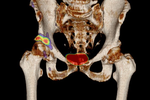
More modern technology may combine the use of a gamma camera (SPECT images) with a CT scanner to obtain two sets of information simultaneously. A combined CT (x-ray) and gamma camera (SPECT) is shown below. The doughnut scanner is the CT and the two square heads are the gamma camera. The bed passes through both scanners in sequence. The combined images are known as SPECT-CT (see image). xSPECT is a new development which takes this concept to a new level. There is more about xSPECT on our website here or for a short slideshow click here.What do we use at Garran Medial Imaging?
At Garran Medical Imaging we have a new Intevo xSPECT system we believe is the most advanced gamma-camera CT fusion systems available in private practice in Australia. This system combines an advanced high resolution gamma camera system with a low dose CT scanner to provide accurate assessments of many different body systems. It can provide direct quantification of tracer uptake which allows a more detailed assessment for many problems. For more on bone quantification click here.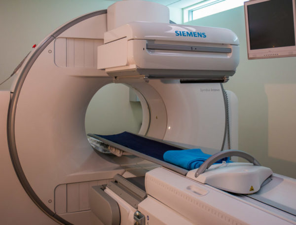
What type of nuclear medicine scans are available?
Using different radiotracers, we complete scans of many different body systems including bone, heart, brain, kidneys, lung, thyroid, among many others. We also provide the entire spectrum of nuclear medicine, including myocardial stress imaging. Some tracers for specific types of disease. A PSMA scan is a scan done for prostate cancer. GMI can do this under a patient specific application as it is currently not easily available in Australia.What are radiotracers and how are they used?
A radiotracer is a substance with a radioactive tag (such as Technetium), which mimics a normal substance in the body to asses different organs or systems. Each radiotracer can show us different body functions and parts. The radiotracer will take time to reach or pass through the area of the body to be examined. The time required is difference for every tracer and system.Are radiotracers dangerour?
Most of the radiotracers are based on the isotope technetium, which has a short (6 hours) half life and is excreted rapidly by the body after your examination. These timing differences determine the specific scheduling of each examination. Radiotracers do involve tiny doses of radiation and do not cause any side effects during their passage through the body. Most of the compounds used are quickly eliminated from the body. The amount of radiation for a single scan is low, and frequently repeated scans are only used when there is a clear benefit to outweigh the slight risks of repeated radiation exposure.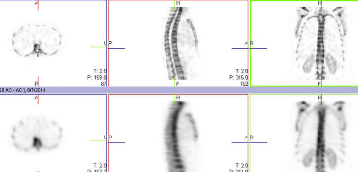
What is the scan procedure?
Many examinations are undertaken in two (or sometimes more) parts. The initial part involves an injection into a vein and sometimes some preliminary pictures. Later more detailed pictures are taken and sometimes a series of images are captures as the tracer passes through the organ system. In some of the test procedures additional activities are undertaken to test the performance of a given organ or system. For example, you may be asked to exercise for cardiac testing. You may also be given a drug to make your kidneys excrete urine or to maximise blood flow to your heart or brain. Sometimes a test is done before and after such an activity.What do the images look like?
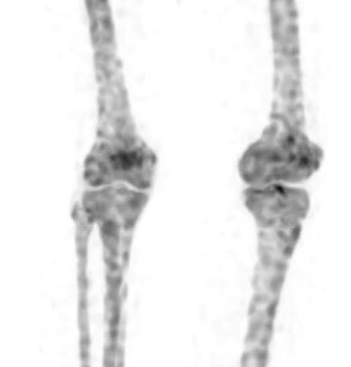
Dr Iain Duncan is one of Australia’s earliest adopters of SPECT-CT, which combines nuclear medicine with CT images. Our bone images show detail that exceeds all other systems Iain has used. This allows us to give you and your doctor more information with lower X-ray.