Quantitative bone imaging is where we measure exactly the amount of tracer in a given volume of interest. This capability has only now become possible with new technological and computer analysis techniques with the use of a specially calibrated xSPECT gamma camera. When analysing the data, we express the result in SUV (standard uptake values), which is a multiple of the expected uptake for the volume assessed. This value is independent of the dose administered and the date of the scan. Higher SUV values indicate more local tracer uptake. This can be invaluable clinically as we can assign more specific significance depending on the magnitude of the values. We can also compare uptake between scans and quantify uptake even in very small lesions.
For example, this image shows bilateral sacroiliitis with SUV maximum values of ~12 which indicates mild to moderate disease. After treatment this scan could be repeated and the specific uptake (in SUV) used to assess response.
Quantitative bone imaging is particularly useful in cancer imaging where both early diagnosis (for example see here) and changes over time are much better evaluated with quantification.
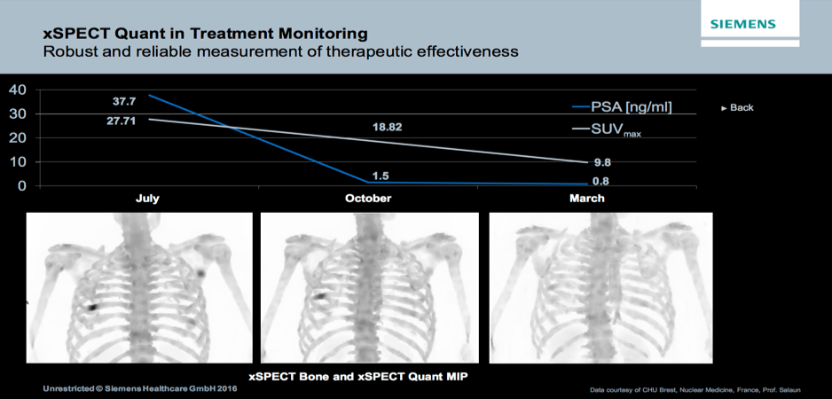
xSPECT quant can be used to assess response to therapy both more precisely and at shorter intervals between scans. This should assist in the delivery of more specific and tailored therapy for many patients. The example shown shows the falling uptake in a rib lesion associated with a fall in PSA which would be interpreted as a good response to therapy.