PSMA Scan for prostate cancer

What is a PSMA scan?

PSMA stands for prostate specific membrane antigen, which is a complex protein found in the cell walls of many types of cell, but greatly increased in prostate cancer. Radio-tracers have been developed to attach to the antigen and thus show up abnormal cells, usually related to prostate cancer. A nuclear (PET or SPECT) scan can detect the radiotracers (Gallium, Technetium or Fluorine) and show their location on a CT scan done simultaneously. At Garran Medical Imaging we do a Technetium scan. We have developed a much higher resolution scan (than standard SPECT-CT) adapting the latest reconstruction techniques and utilising specialised windowing. Our results are comparable with Gallium PET scans.

What is it used for?

A PMSA scan is used  either in the diagnosis or follow-up of prostate cancer. It can show cancer activity in the prostate , surrounding tissue, lymph glands, or bone. It can help assess cancer spread and response to therapy. It can help you and your doctor decide on the best treatment options.

Who should have a PSMA scan?

Currently the exact role of the scan is in development as the technology is new. You need to discuss this with your treating doctor or surgeon (who can contact us if required). The Tc-PSMA scan requires a patient by patient application (which we can arrange), since we import the tracer for each patient on a case by case basis. It comes from Germany and it is not yet approved by our TGA (Therapeutic Goods Administration). Nevertheless we feel it is completely safe to use and have already used it extensively.

What is the PSMA scan procedure?

The scan is  done much like any other nuclear scan and requires an injection of a radio-isotope tracer followed by a series of scans, usually ~3-4hrs after the injection. The scan procedure itself will probably take 30-45 minutes. More detail about nuclear scans can be found here and the actual procedure here. For patients who have previously had a prostatectomy we also do a second scan the day after (24hr scan) as this gives us a much better evaluation of the prostate bed and around the bladder.

If I have prostate cancer is this the only scan I need?

No. The this can outline the extent of the cancer you often need other scans to better detail within and immediately around the prostate, such as an MRI scan. A bone scan and CT scan are often also done for  treatment planning or treatment followup.

What do the images look like?

The scans produced are both a whole body overview scan and a 400 slice hybrid nuclear-CT scan which show your entire body in detail. Key images are produced by the reporting physician for easy review by your doctor. Examples of key images are shown below.

Can I be sure GMI is producing clinically useful results?     
Yes. See here.

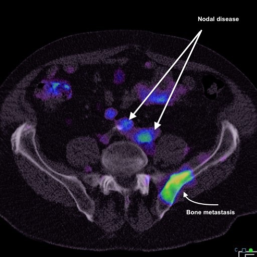
PSMA scan showing two abnormal lymph nodes and metastatic disease in the iliac bone

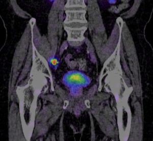
Scan showing prostate cancer in a lymph node. The uptake in the middle is tracer in the bladder

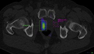
This shows cancer in the prostate gland