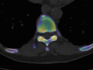
A mobile friendly slideshow of this post about xSPECT bone can be found here. For those with a professional interest see our paper in the European Journal of Hybrid Imaging , see Dr Iain Duncan’s lecture at the 2019 World Molecular Imaging Summit at Lausanne in Switzerland, or visit Siemens healthineers.
xSPECT Bone
NUCLEAR IMAGING: THE OLD V NEW
Nuclear Medicine has made a dramatic transformation between “the old” and “the new” over the last decade (see my post Nuclear Medicine = New Clear Medicine). The fusing together nuclear medicine and CT scans was given the description SPECT/CT. SPECT is short for “single photon emission computerised tomography” and represents cross sectional nuclear medicine, but really no one outside the field understands the acronym. The CT part represents the much better know computerised tomography or “CAT” scan. In essence SPECT/CT indicates the fusion of anatomic x-ray and functional nuclear medicine.
Though this was a giant leap forward there was still a spatial limitation because of the SPECT component of the fused imaging. This limitation was considered fundamental as nuclear medicine is based on a low energy gamma wave. This gamma wave is generated by the tracer inside a subject/patient inside compared with an x-ray being fired in a parallel beam from outside. The gamma rays don’t come in a straight line from a single source -thus a fundamental physical limitation of spatial accuracy. Siemens however came up with an ingenious solution by using the detailed spatial information about the tissues from the CT scan (x-rays) to reinterpret/reintegrate the gamma ray (SPECT) data arriving from within the patient.
HOW XSPECT BONE WORKS
The “reconstruction algorithm” reconfigures the gamma ray information by combing the gamma-ray and x-ray data at a fundamental level. This substantially improves the spatial accuracy and detail of the SPECT part of the image. The next paragraph explains more but can be skipped…
In their publications Siemens notes “Using CT-based segmentation in the reconstruction, xSPECT Bone achieves a sharper definition of bony margins and lesion boundaries. The resulting reconstructed SPECT images (xSPECT Bone) appear to truly reflect the distribution of metabolically active bone within the skeletal system with sharper differentiation between cortical and spongy bone in the vertebrae, as well as in flat bones like the pelvis, scapula and skull. This also provides fine detail which helps differentiate between the cortex and marrow cavity in long bones and improved separation of bone from joint spaces—both for large joints such as the knee and shoulder and for small bones such as carpals and tarsal bones.”
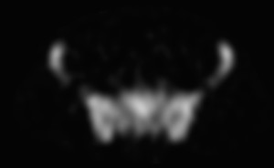
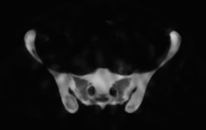
The end result is a high resolution SPECT bone scan when compared with standard SPECT. Everyone can appreciate the difference as shown below. Fig 1 and 2 compare the conventional SPECT bone reconstruction (Fig 1) versus the xSPECT bone reconstruction (Fig2). Both images are taken from a single slice of the human pelvis and (images courtesy of Garran Medical Imaging).
Figs 1-2 below.
xSPECT single slice PELVIS
This advance has side stepped the physical limitation of gamma-ray imaging and the bone uptake resolution is taken to a new level.
This is further illustrated using image phantoms as in Fig.3 below. These images were performed on our equipment at Garran Medical Imaging and are courtesy of Jason Tse1, Farshid Salehzahi1, and Nick Ingold2 .

Fig3. This is a phantom to test the resolution of our camera. The image on the left shows xSPECT technology and on the right using SPECT reconstruction
WHAT DOES XSPECT BONE MEAN FOR THE PATIENT AND REFERRING DOCTOR?
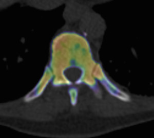
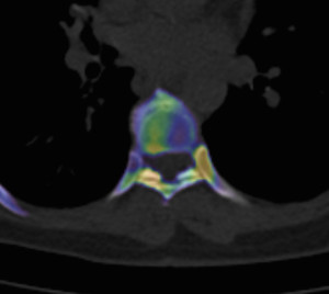
The cases below all show bone isotope uptake in some very small locations that were previously rarely identified on SPECT-CT (again images courtesy Garran Medical Imaging).
Our first 200 cases have shown us that xSPECT imaging makes a BIG difference, giving additional information in in 71% of scans and changing the diagnosis in 20%, when compared to the current best SPECT scans. For the whole study see our research in the European Journal of Hybrid Imaging.
All of these xSPECT-CT scans (Figs 3-5 above) show abnormalities that could not be localised with conventional SPECT-CT alone.
Dr Iain Duncan has been a pioneer in the application of this technology and was recently invited to share his experience with others at a World Molecular Imaging Summit in Lausanne, Switzerland, May 2019 (see his lecture here).
For more about xSPECT bone see Dr Duncan’s research.
See our video with a 1minute UPDATE on Molecular Imaging without compromise.
